Osteora
Choosing Titanium Mesh For Cranial Repair in 2026 requires more than comparing prices or looking at product photos. The right option depends on the defect’s size and location, the patient’s anatomy, the planned fixation method, and the surgeon’s assessment of protection and contour. A mesh that looks suitable on a screen may need adjustment when matched to a real skull defect. Small details matter: edge position, screw placement, implant thickness, and how the reconstruction will appear on follow-up imaging.
This guide examines material specifications, design options, fit, handling, and questions to raise with the surgical team. It also distinguishes general product information from decisions that require clinical judgment. No single mesh design is right for every patient. Even a well-planned reconstruction can involve trade-offs, and those should be discussed rather than glossed over. A verified quotation from a named cranial-repair specialist was not provided, so I have not invented or misattributed one. Add an expert’s exact, source-checked statement before publication to strengthen the article’s authority and give readers a useful professional perspective.
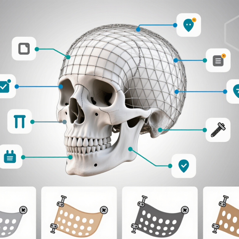
Assess the defect from current CT images and operative records, not from a skull measurement alone. Note its dimensions, depth, location, and curvature. A broad frontal opening may need a different contour from a small defect near the orbit. Measure twice. Check whether the dura, venous sinuses, or nearby nerves affect safe fixation and mesh placement.
Then consider the patient’s clinical needs. Prior infection, previous surgery, thin scalp tissue, and expected healing can influence implant design and coverage plans. A low-profile mesh may reduce prominence under delicate tissue, but it still needs adequate strength and secure fixation. Ask about planned imaging and other procedures; titanium can create image artifacts, and compatibility depends on the specific implant and scanner conditions.
Age and activity matter, too. A growing child requires different planning from an adult with a stable defect. Review the mesh’s pore size, thickness, contourability, and screw options against the defect and the surgeon’s technique. Small details count. The best fit on a model may not sit perfectly once exposed tissue and real bone edges are considered. Discuss these uncertainties with the cranial surgeon and, when needed, radiology and reconstructive specialists.
Titanium mesh selection is a balance between coverage, contour, and handling. Perforated sheets allow tissue access and fluid passage; woven or more open designs can feel flexible but may be harder to contour evenly. Patient-specific meshes can match a defect closely, yet accurate imaging and planning matter. Fit matters. A poorly seated edge can irritate soft tissue, even when the material itself is suitable.
Thickness affects stiffness, shaping, and profile beneath the scalp. Thinner mesh may sit closer to the bone, but it is not automatically the best choice for every defect. ASTM F67 gives minimum tensile strengths of 240 MPa for Grade 1 commercially pure titanium and 345 MPa for Grade 2. These figures describe the material specification, not the strength of a finished mesh or a patient’s outcome. Small detail. Compare pore size, edge finish, and surface texture alongside thickness. A smooth surface may ease tissue contact, while a rougher finish can change handling and integration considerations. I would not treat one surface design as universally superior. Published clinical reports vary in patients, defect sizes, and follow-up, so ask the surgical team how the chosen design fits the specific anatomy and closure plan.
How to Choose Titanium Mesh for Cranial Repair in 2026?
Evaluate the defect on thin-slice CT, including curvature, bone thickness, and nearby sinuses. A mesh that looks accurate on a screen may still lift at the edge. Small gaps matter. Check that the planned contour sits flush without pressing on the scalp or leaving sharp margins. A 2016 systematic review by Malcolm and colleagues in Neurosurgery reported an overall cranioplasty complication rate of 19.5% across 1,093 patients. This figure covers different materials and complications; it is not a titanium-mesh-specific risk estimate.
Fixation should match the available bone, not just the drawing. Map screw positions around thin bone and avoid locations where purchase may be unreliable. Low-profile fixation can reduce prominence, but the best choice depends on access and local anatomy. Access changes everything. Plan the incision and exposure with the surgical team: limited exposure may make contouring and secure fixation harder, while wider exposure can affect soft-tissue coverage. Consider how the mesh will be handled through the opening, and whether edges can be inspected before closure. Digital planning helps, but it cannot fully predict scalp tension or intraoperative fit. That uncertainty deserves a final hands-on check.
Biocompatibility is more than a material label. Confirm the mesh uses implant-grade titanium, with traceable material records and documented manufacturing controls. Ask how surface finishing, cleaning, and sterilization are verified; residues or rough edges can matter. A certificate alone is not the whole story. This step is easy to overlook.
Imaging needs should be discussed before selection. Titanium can still create artifacts, and their extent depends on mesh thickness, shape, and imaging method. Ask the clinical team whether postoperative CT detail is important near the repair site. For MRI, check the device’s labeling and conditions rather than assuming every titanium implant is suitable for every scan. Not all meshes behave alike.
Check which standards apply to the specific device and intended market. Depending on the material and product, relevant references may include ISO 10993 for biological evaluation, the ISO 5832 series for metallic implant materials, or applicable ASTM material specifications. These references are not interchangeable, and the right set depends on design and regulatory context. Request current test reports, material specifications, and sterilization documentation from the supplier. If evidence is incomplete, pause and ask a qualified clinician or materials specialist to review it. Safety is not a guess.
Planning titanium mesh placement begins with thin-slice CT and a clear map of the defect, nearby venous sinuses, and the intended fixation points. Review the scalp condition and soft-tissue coverage; a prominent edge beneath thin tissue may later become an exposure problem. Small gaps matter. Shape the mesh to restore contour without pressing on the dura, and check that screws can be placed safely. A 2015 systematic review in Neurosurgical Review reported a pooled infection rate of 7.7% across cranioplasty studies. That figure covers varied materials and patients, so it should not be treated as a titanium-mesh-specific prediction.
Before surgery, document neurological status and any existing wound concerns. After placement, use the treating team’s imaging plan to check implant position and establish a comparison point. Monitoring should include the incision, scalp tension, pain, temperature, and neurological changes. Watch the skin. New swelling, drainage, increasing redness, fever, worsening headache, confusion, or weakness needs prompt clinical assessment. The review’s complication data reinforce why follow-up should be individualized, especially when tissue coverage is limited or the patient has had prior infection. A monitoring plan can still miss subtle changes; caregivers should know whom to contact and what changes to report.
Perforated sheets allow tissue access and fluid passage. Woven or open designs may feel more flexible, but can be harder to contour evenly.
No. Thinner mesh may sit closer to the bone, but stiffness and shaping needs vary by defect. Fit matters.
Compare pore size, edge finish, and surface texture. Smooth surfaces may ease tissue contact, while rougher finishes can affect handling.
No. The cited minimums are 240 MPa for Grade 1 and 345 MPa for Grade 2 titanium. They describe material specifications, not a finished mesh’s strength or a patient’s outcome.
Ask for traceable material records and documented manufacturing controls. Check how finishing, cleaning, and sterilization are verified. A certificate alone is not the whole story.
Yes. Titanium can create imaging artifacts, and their extent depends on mesh thickness, shape, and imaging method. Ask whether CT detail near the repair site matters.
Do not assume so. Check the specific device labeling and scan conditions with the clinical team. Not all meshes behave alike.
Depending on the device, relevant references may include ISO 10993, the ISO 5832 series, or applicable ASTM specifications. Request current test reports and sterilization records. The right set depends on design and regulatory context.
A poorly seated edge can irritate soft tissue, even when the material is suitable. Patient-specific designs may match a defect closely, but accurate imaging and planning matter. I may be overlooking a detail, so the surgical team should review the fit.
Choosing Titanium Mesh For Cranial Repair in 2026 begins with understanding the defect’s size, location, shape, and the patient’s clinical needs. These factors help guide the selection of mesh type, thickness, and surface design, balancing structural support with contour and handling requirements. The implant should be evaluated for how accurately it can fit the defect, which fixation options are suitable, and whether the surgical approach provides practical access for placement.
Planning should also account for biocompatibility, expected imaging needs, and applicable technical standards. Before surgery, the team can review implant positioning and fixation to support a stable, carefully planned repair. After placement, postoperative monitoring helps assess healing, implant position, and any changes that may require clinical attention. A thoughtful process that connects patient assessment, material selection, surgical planning, and follow-up can help clinicians make informed choices for cranial reconstruction.